 |
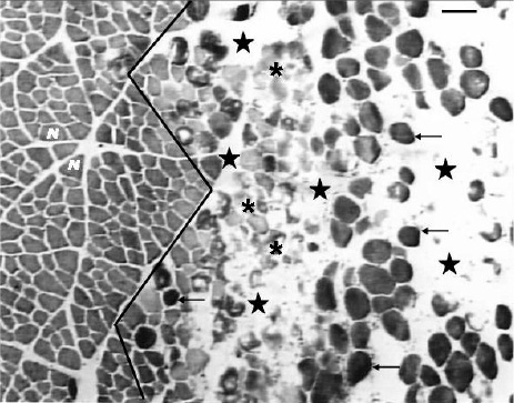
Figure 1.
Cross-section of tibialis anterior (TA) muscle middle belly, 4½ hours after induced injury. Note two different muscle regions separated by a black continuous line: one composed by normal muscle fibres (N, left side of the line), and the other one composed by injured fibres (right side of the line). Hypercontracted fibres (arrows), disrupted fibres (asterisk) and large clear areas among the muscle fibres (stars) characterize the injured area, indicating edema and muscle fibres disruption. Bar: 40 µm. Toluidine blue stain.