 |
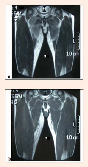
Figure 1.
Coronal T2-weighted image (a) shows high signal intensity in the bilateral adductor magnus with slight swelling and subcutaneous edema at the medial thigh. Coronal T1-weighted image (b) shows an iso-signal intensity compared with other muscles.