 |
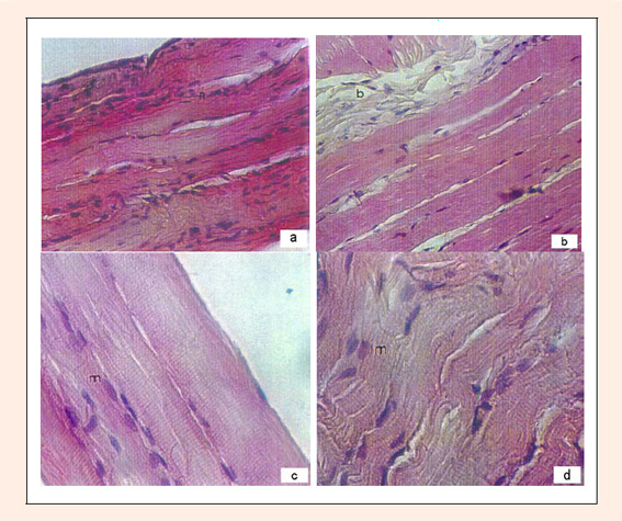
Figure 3.
Histological appearance of the skeletal muscle in rat. (a) The tissue sample belonging to CON group (no Cr supplementation) diaphragm muscle in rat (haematoxylin-eosin (HE)-stained section, n = nukleous, original magnification X20). (b) The histological changes appear in the diaphragm muscle tissue sample of the INCO group. (HE-stained section, original magnification X20, fibroblast and collagen fibers are seen with increased connective tissue between muscle cells = b). (c) The myoblasts are seen between hypertrophic gastrocnemius muscle cells in the tissue sample belonging to CREAT-I group (Cr supplementation 1 g·kg·day) (HE-stained section, original magnification X40, myoblast = m). (d) The tissue sample belonging to CREAT-II group (Cr supplementation 2 g·kg·day) diaphragm muscle in rat (HE-stained section, original magnification X40, the round-shaped myoblasts are seen between the hypertrophic muscle cells = m).