 |
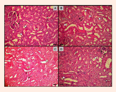
Figure 4.
Representative photomicrographs of kidney from Normal diet Sedentary (SED), Creatine diet Sedentary (CRESED), Normal diet Exercised (EXE), Creatine diet Exercised (CREEXE) groups after 4 weeks of supplementation. Magnification 400x. Scale bar = 25µm. The SED group (panel A) had renal corpuscles with a thin contour and well defined Bowman capsules (continuous arrows). Glomerular capilar (dotted arrow) were not congested and had intra-capsular spaces preserved. Renal tubules showed preserved cellular morphology (cuboid epithelium) and distinct lumen, plus proximal tubule convoluted (stars). In CRESED group (panel B), possible renal damage morphology was verified: renal corpuscles with outline of difficult delimitation and irregularity of the Bowman´s Capsule (continuous arrow), dilation of capillary glomeruli (dotted arrow) and intra-capsular space not preserved. Renal Tubules exhibited cellular morphologic regularity (cuboid epithelium), but indefinite space tubular (arrowsheads). Panels A, C and D, show kidney (cortex part) with preserved renal morphology: Renal corpuscles with very defined outline and regularity of the Bowman´s Capsule (continuous arrow), no dilation of glomeruli capillary (dotted arrow) and intra-capsular space preserved. Renal Tubules exhibited cellular morphologic regularity (cuboid epithelium), and definite tubular space (arrowheads).