 |
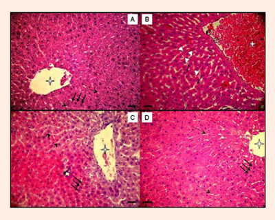
Figure 5.
Representative photomicrographs of liver from Normal diet Sedentary (SED), Creatine diet Sedentary (CRESED), Normal diet Exercised (EXE), Creatine diet Exercised (CREEXE) groups after 8 weeks of supplementation. Magnification 400x. Scale bar = 25µm. The SED group (panel A) had a central vein which was uncongested (star), normal hepatocytes (continuous arrows), and slight capillary sinusoids (dotted arrows). The hepatocytes show standard morphology with a big and centralized nucleus. In CRE group (panel B) possible hepatic damage morphology was verified: congested central vein (star) and with some polymorphonuclear in the peripheral position (circular delimitations), walls of the sinusoids showed numerous Kupffer cells (arrowheads) and with swelling lumen by the blood cells (star). The EXE group (panel C) showed hepatocytes (continuous arrows) distributed throughout interconnected plates from the central vein (star) and separated by slight capillary sinusoids (dotted arrows). The EXECRE group (panel D) had preserved hepatic morphology: central vein with no congested (star), hepatocytes (dotted arrows) correctly arranged in trabecules running radiantly from the central vein and separated by thin sinusoids (continuous arrows). They were regular and contained a large spheroid nucleus.