 |
Figure 1.
Typical MRI images showing transverse sections of the mid-thigh taken before (pre) and after (post) 8 wk of cycle training with BFR. The images show identical sections midway along the femur in the same subject (KK).
|
|
|
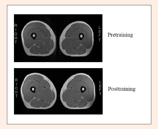
Figure 1.
Typical MRI images showing transverse sections of the mid-thigh taken before (pre) and after (post) 8 wk of cycle training with BFR. The images show identical sections midway along the femur in the same subject (KK).
|