 |
Figure 2.
The 3D tibiofemoral model was simultaneously matched to its corresponding two 2D images on biplane radiographs at extension, 15°, 30°, 60°, 90°, and 120°. The spherical marker was indicated in white
|
|
|
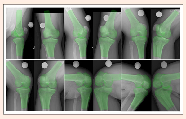
Figure 2.
The 3D tibiofemoral model was simultaneously matched to its corresponding two 2D images on biplane radiographs at extension, 15°, 30°, 60°, 90°, and 120°. The spherical marker was indicated in white
|