 |
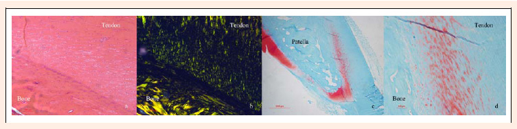
Figure 2.
A sagittal section of the patella-patellar tendon junction of the CON group. The H&E staining of a section from the CON group shows good alignment of the cell profile and a clear tidemark (arrow) (A). The polarized light image revealed well-aligned collagen fibers, and a dark band was found in the fibrocartilage zone (B). The proteoglycan profile located in the fibrocartilage zone was even and continuous (C, D) (100× for a, b, and d; 20× for c).