 |
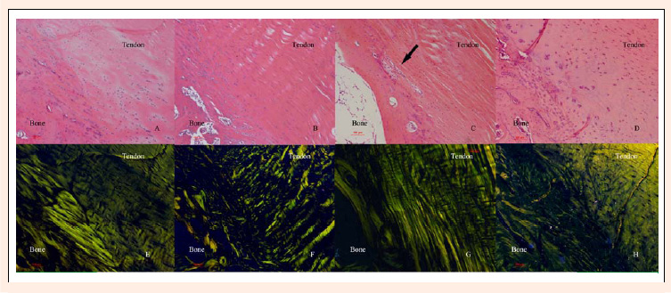
Figure 3.
The histological characteristics of post-injury in BTJ. Compared with the CON group, the ND1W group exhibited decreased cell density, an unclear tidemark, and some scar tissue (A). The collagen alignment was normal, but the waviness of the collagen fibers was diminished (E). The ND2W group showed a poor cell profile, no detectable tidemark, and a different collagen fiber diameter (B, F). The cell profile in the ND4W group was poor, and the chondrocytes proliferated in the fibrocartilage zone. A chondrocyte island formed (arrow), and the collagen fiber diameter increased; the waviness decreased (C, G). The ND8W group showed a poor cell profile, decreased cell density, scar tissue formation, and diminished waviness of the collagen fibers (D, H). (100 ×).