 |
Figure 1.
Ultrasound images of Vastus Lateralis (VL) and Intermedius (VI) muscles. Left: cross-sectional image. Right: longitudinal image representing pennation angle (α) and fascicle length (L) of VL.
|
|
|
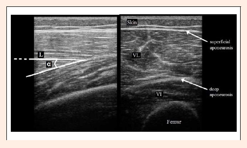
Figure 1.
Ultrasound images of Vastus Lateralis (VL) and Intermedius (VI) muscles. Left: cross-sectional image. Right: longitudinal image representing pennation angle (α) and fascicle length (L) of VL.
|