 |
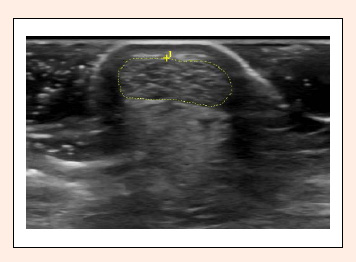
Figure 2.
Outline of the Achilles tendon (AT) cross-sectional area (CSA). Internal software of the GE Logic was used. This image was taken in the transverse plane at the thinnest portion of the tendon between the calcaneus and gastrocnemius insertion sites. The skin can be seen at the top of the image in a white semicircular arch around the posterior portion of the tendon. Photo: A. Wayne Johnson