 |
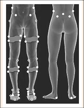
Figure 1.
Rendering of lower-extremity marker placement. Static (gray-filled) markers were removed after the standing calibration. Dynamic (white-filled) markers were kept on during the running trials. Static markers were placed on the L5/S1 joint space, and the bilateral iliac crests, anterior and posterior superior iliac spines, greater trochanters, lateral and medial knee joint spaces, lateral and medial malleoli, distal second (foot) phalanges, base of the 1st and 5th metatarsals, and calcanei.