 |
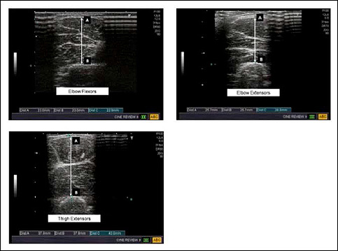
Figure 1.
Typical example of ultrasound images obtained in three muscular sites. Panels A and B represent adipose/muscle interface and muscle/bone interface, respectively; arrow represents the measured thicknesseses.
|
|
|
Figure 1.
Typical example of ultrasound images obtained in three muscular sites. Panels A and B represent adipose/muscle interface and muscle/bone interface, respectively; arrow represents the measured thicknesseses.
|