 |
Figure 3.
Scar dimensions (upper graph) and pennation angle of superficial, deeper and “typical” fascicles of the biceps femoris (lower graph) during the intervention period.
|
|
|
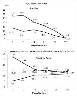
Figure 3.
Scar dimensions (upper graph) and pennation angle of superficial, deeper and “typical” fascicles of the biceps femoris (lower graph) during the intervention period.
|