 |
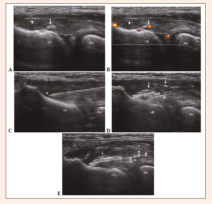
Figure 1.
Ultrasonographic findings of the common extensor tendon in a 44-year-old woman during PRP injection
A. Longitudinal image showing amorphous hyperechoic calcification (arrow) and a partial-thickness tear (arrow head) in the common extensor tendon. B. Longitudinal power Doppler image showing increased vascularity around a calcification (arrow) and partial-thickness tear (arrow head) in the common extensor tendon. C. Longitudinal image showing the needle tip (star) located in the area of a partial-thickness tear. D. Post-injection ultrasonographic image showing injected PRP (arrow) spread within the tendon; the star indicates the injected needle. E. Image 5 minutes post-injection showing some of the injected PRP remaining (arrow) near the injection site (star) and the rest extending up to 1.72 mm (arrowhead) beyond injection site surrounding soft tissues. LE: lateral epicondyle of the humerus, RH: radius head.
A. Longitudinal image showing amorphous hyperechoic calcification (arrow) and a partial-thickness tear (arrow head) in the common extensor tendon. B. Longitudinal power Doppler image showing increased vascularity around a calcification (arrow) and partial-thickness tear (arrow head) in the common extensor tendon. C. Longitudinal image showing the needle tip (star) located in the area of a partial-thickness tear. D. Post-injection ultrasonographic image showing injected PRP (arrow) spread within the tendon; the star indicates the injected needle. E. Image 5 minutes post-injection showing some of the injected PRP remaining (arrow) near the injection site (star) and the rest extending up to 1.72 mm (arrowhead) beyond injection site surrounding soft tissues. LE: lateral epicondyle of the humerus, RH: radius head.