 |
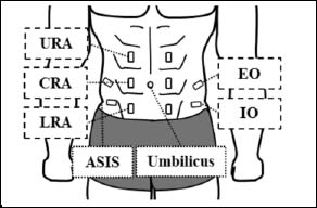
Figure 2.
Schematic illustration of the electrode placements in the upper, central, and lower portions of the rectus abdominis (URA, CRA, and LRA, respectively) and the external and internal obliques (EO and IO, respectively). ASIS, anterosuperior iliac spine.