 |
Figure 2.
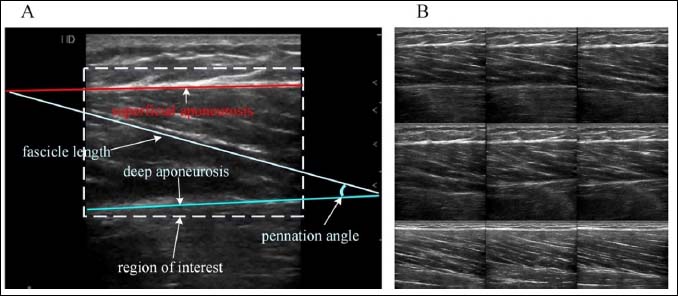
Ultrasound image showing the region of interest, fascicle length, superficial and deep aponeuroses, and pennation angle (A). Ultrasound imaging screenshots during running (B).
|
|
|
Figure 2.
Ultrasound image showing the region of interest, fascicle length, superficial and deep aponeuroses, and pennation angle (A). Ultrasound imaging screenshots during running (B).
|