 |
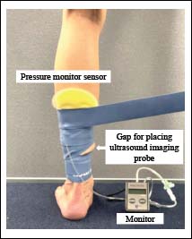
Figure 3.
Illustration of wrapping tissue flossing and inserting pressure monitor sensor. The floss band was wrapped around the non-dominant calf from just above the most prominent part of the medial ankle to the inferior border of the patella, extending 1.5 times the natural length and overlapping the bands by 50%. The Kikuhime pressure monitor sensor was placed at the largest bulge at the center of the posterior calf.