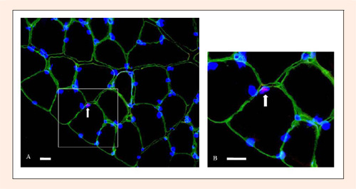
Figure 1. Identification of satellite cell and myonuclei. (A) A satellite cell was detected with triple staining for Pax7(red), myonuclei(blue) and laminin(green). (B) An area is viewed at a higher magnification. The arrow is showing a satellite cell. Bar = 20µm.