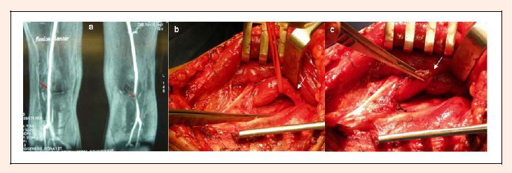
Figure 1. a) Functional magnetic resonance angiography (fMRA) with forced plantar flexion, showing bilateral arteriovenous collapse, most evident in the left lower extremity (red arrows). No anatomic abnormality of muscle or ligament insertion. b) Surgical exposure with posterior approach shows a hypertrophied plantaris muscle (with red vessel loop) compressing the vascular bundle (white arrows). c) Section of the hypertrophied plantaris muscle and neurovascular release of adhesions (white arrows).