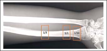
Figure 1. Representative scan of the distal radius. The three analyzed regions (boxed insets) 1/3, 1/6, and 1/10 of the bone length from the distal end of the radius. Measurements were performed using the software supplied by the manufacturer (DCS-3000, Software Ver. 5.0, Aloka, Japan).