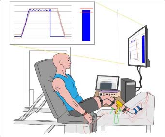
Figure 1. Experimental setup. The force exerted by the dorsiflexor muscle of the dominant leg was measured with a force transducer attached under the foot. Participants were asked to minimize coactivation of thigh and trunk muscles. High-density electromyography (HD-EMG) signals were recorded from the tibialis anterior muscle of the dominant leg with a semi-resistant adhesive grid (yellow pad). Surface EMG recordings were also obtained from a pair of surface electrodes placed over soleus and gastrocnemius medialis (green wires). The reference electrodes were placed at the wrist for the bipolar recordings and at the ankle for the grid (red wires). Two goniometers were placed over the knee and the ankle joints to measure joint angle. Visual feedback was provided on a monitor of the target force (red lines) and the applied force (blue lines) during the ramp-up, plateau, and ramp-down phases (middle screen) and on a moment-to-moment basis (right side of screen). The display covered approximately 80% of the screen.