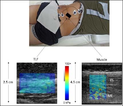
Figure 1. The experimental set-up. The ultrasound probe was placed 2.5 cm away from the L3 spinous process at the L3-L4 level to visualize the thoracolumbar fascia (TLF), the erector spinae (ES) and deep multifidus (MF). Bipolar electrodes were also placed on the ES and MF. TLF modulus was estimated by taking several circular regions of interest inside a rectangular coded box of 1.5. x 2 cm and ES and MF moduli were determined separately by drawing smaller circles within a 4 x 3 cm rectangular box. The color scale was extracted from the software and is enlarged so that the measurement scale is easily visible.