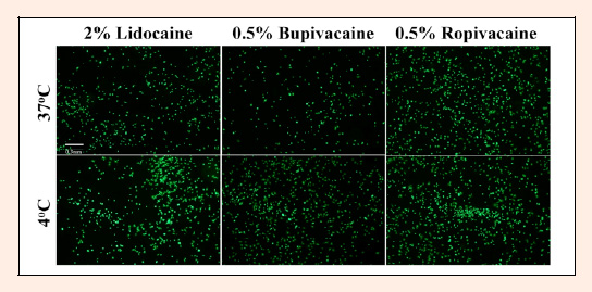
Figure 2. Viable chondrocytes stained with Calcein AM, visualized at 5x magnification under a FITC filter. Chondrocytes were treated with 2% lidocaine, 0.5% bupivacaine, or 0.5% ropivacaine and incubated at 37°C or 4°C. Briefly, when chondrocytes are treated with lidocaine or bupivacaine, there appears to be a higher density of viable chondrocytes when incubated at 4°C compared to chondrocytes incubated at 37°C. Viable cell density appears to remain constant for chondrocytes treated with ropivacaine across both 37°C and 4°C. Results from fluorescent microscopy appears to be consistent with results from PrestoBlue® fluorescence assay. Scale bar = 0.3mm.