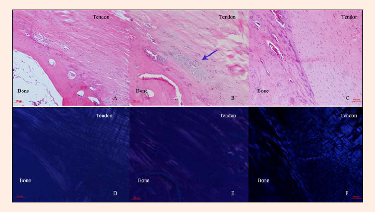
Figure 3. A week 4 representative sagittal section of the patella-patellar tendon junction after injury and training. H&E-stained sections from the AI (A) and PIERT (B) groups indicated an indistinct cell profile, lowered cell density, and unclear tidemark, but the CON group (C) presented well-aligned collagen fibres and a clear tidemark. Polarised microscopy images demonstrated poorer collagen alignment in the AI group (D) and PIERT group (E) than in the CON group (F).