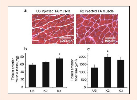
Figure 3. Muscular hypertrophy of tibialis anterior induced by knockdown plasmids. At 2 weeks after introduction of knockdown plasmids, the tibialis anterior was dissected out and weighed (n=10 per group). a Representative micorograph of tibialis anterior injected with U6 (left) and K2 (right) plasmids. Centrally located nuclei caused by plasmid injection with electroporation are apparent in both micrographs. b Muscle mass was increased by knockdown plasmid introduction. c Muscle fiber size (cross-sectional area) was measured for 1000 fibers per sample, and this was also shown to be increased by introduction of the knockdown plasmid. * p < 0.05 vs U6 control.