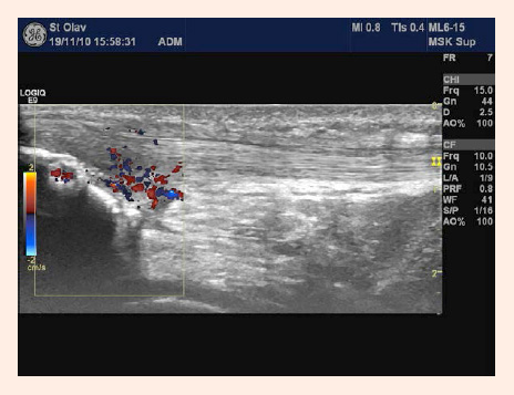
Figure 3.
Neovascularization in the proximal part of the patellar tendon visualized with colour flow imaging (Photo by Salvesen ES).