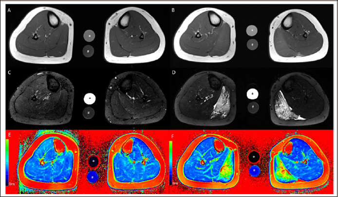
Figure 3. Axial T1-weighted (A, B), axial T2-weighted TIRM (C, D) and axial T2 mapping (E, F) images of the lower leg at baseline (A, C, E) and 60 h after standardized eccentric exercise of the calf muscles (B, D, F). Muscle edema is apparent in the medial gastrocnemius muscle in the post-exercise images (B, D, F). This participant wore the compression sock on the left calf for 60 h after eccentric exercise. The calibration tubes contain 40 mmol/L NaCl (*) and 40 mmol/L NaCl 5% agarose (#).