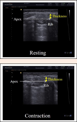
Figure 3. Ultrasound images of the serratus anterior muscle at rest and during MVIC. The rib was used as a reference for measurement of the serratus anterior. The vertical yellow lines, spaced out to incorporate the apex of the rib, were drawn from the rib to the superior fascial border of the serratus anterior. MVIC: maximal voluntary isometric contraction.