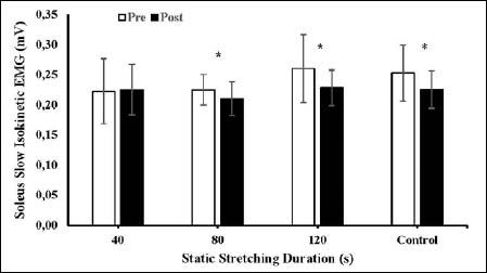
Figure 3.
Illustrates the interaction of conditions (antagonist stretching for 40-s, 80-s, 120-s and control) and time (pre- and post-test) for soleus slow isokinetic EMG. * indicates significance.