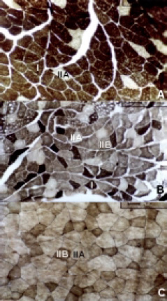
Figure 4. Reprasantative examples of transverse sections (10µm) of soleus (A), extensor digitorum longus - EDL (B) and gastrocnemius - white portion muscles (C) from sedentary group with no supplementation (CON), stained for myofibrilar ATPase activity after acid pre-incubation at pH 4.6. Labels provided for type I (slow), IIA (intermediary) and IIB (fast). Magnify of 200X.