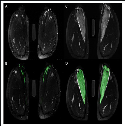
Figure 4. Volume of edematous muscle was assessed in the T2-weighted TIRM images acquired 60 h after standardized eccentric exercise of the calf muscles (C, D). Exemplary images before (C) and after color-coded manual segmentation (D) of the edematous medial gastrocnemius muscle are shown. Hyperintense vessels were segmented in the baseline images (B) and subtracted from the segmented volume in the post-exercise images (D).