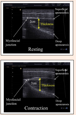
Figure 4. Ultrasound images of the supraspinatus muscle at rest and during MVIC. The point of 2 cm from the angle formed by the superior muscular fascia of the supraspinatus was measured. Yellow lines represent the method for off-line supraspinatus muscle thickness measurement. MVIC: maximal voluntary isometric contraction.