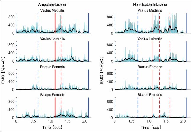
Figure 4. Temporal pattern of EMG of vastus medialis (VM), vastus lateralis (VL), rectus femoris (RF), and long head of biceps femoris (BF) at one typical turn cycle. The light blue lines show the rectified EMG signal, and black lines show the smoothed rectified EMG signal.