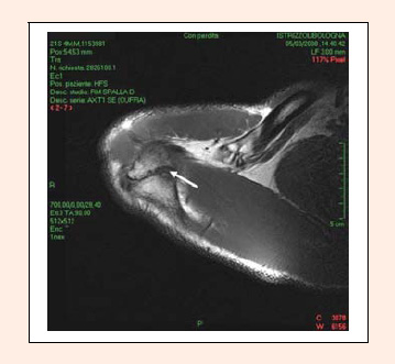
Figure 5.
T2-weighted axial MRI scan examination shows clearly an unfused acromial apophysis with oedema around the synchondrosis site.