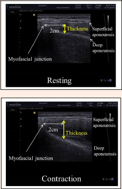
Figure 5. Ultrasound images of the infraspinatus muscle at rest and during MVIC. The ultrasound images were taken from the inferior-most aspect of the superficial fascia to the most superior aspect of the infraspinous fossa, which appeared as a bright, continuous hyperechoic line spanning the width of the screen.MVIC: maximal voluntary isometric contraction.