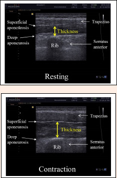
Figure 6. Ultrasound images of the rhomboid major muscle at rest and during MVIC. Ultrasound images of the rhomboid major muscle located between the trapezius and posterior serratus muscles. The thickness can be examined as the vertical length between the inferior echogenic fascial line of the trapezius and the superior line of the posterior serratus. MVIC: maximal voluntary isometric contraction.