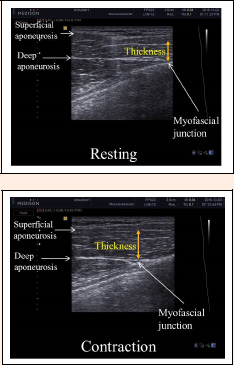
Figure 7.
Ultrasound images of the pectoralis major muscle at rest and during MVIC. Thickness was measured at each aspect of distance between superficial and deep aponeurosis. MVIC: maximal voluntary isometric contraction.